 | Info
Sheets |
| | | | | | | | | | | | | | | | | | | | | | | | |
 | Out-
side |
| | | | |
|
| | | | |
Result : Searchterm 'SCan Time' found in 1 term [ ] and 48 definitions [ ] and 48 definitions [ ] ]
| | previous 41 - 45 (of 49) nextResult Pages : [1] [2 3 4 5 6 7 8 9 10] |  | |  | Searchterm 'SCan Time' was also found in the following services: | | | | |
| |  |
| |
|
SPEEDER™ is Toshiba's patented parallel imaging technology for high-speed imaging. This technique reduces examination time for MRI studies by a factor of up to three while enabling clinicians to capture high-quality images with detailed diagnostic information. With SPEEDER™, cardiac and MRA examinations are more patient-friendly and tolerable with shorter scan times and improved image quality.
See also Sensitivity Encoding. | | | | | |
| | | | | |
| |
|
(SENSE) A MRI technique for relevant scan time reduction. The spatial information related to the coils of a receiver array are utilized for reducing conventional Fourier encoding. In principle, SENSE can be applied to any imaging sequence and k-space trajectories. However, it is particularly feasible for Cartesian sampling schemes. In 2D Fourier imaging with common Cartesian sampling of k-space sensitivity encoding by means of a receiver array enables to reduce the number of Fourier encoding steps.
SENSE reconstruction without artifacts relies on accurate knowledge of the individual coil sensitivities. For sensitivity assessment, low-resolution, fully Fourier-encoded reference images are required, obtained with each array element and with a body coil.
The major negative point of parallel imaging techniques is that they diminish SNR in proportion to the numbers of reduction factors.
R is the factor by which the number of k-space samples is reduced. In standard Fourier imaging reducing the sampling density results in the reduction of the FOV, causing aliasing. In fact, SENSE reconstruction in the Cartesian case is efficiently performed by first creating one such aliased image for each array element using discrete Fourier transformation (DFT).
The next step then is to create a full-FOV image from the set of intermediate images. To achieve this one must undo the signal superposition underlying the fold-over effect. That is, for each pixel in the reduced FOV the signal contributions from a number of positions in the full FOV need to be separated. These positions form a Cartesian grid corresponding to the size of the reduced FOV.
The advantages are especially true for contrast-enhanced MR imaging such as
dynamic liver MRI (liver imaging) ,
3 dimensional magnetic resonance angiography (3D MRA), and magnetic resonance cholangiopancreaticography ( MRCP).
The excellent scan speed of SENSE allows for acquisition of two separate sets of hepatic MR images within the time regarded as the hepatic arterial-phase (double arterial-phase technique) as well as that of multidetector CT.
SENSE can also increase the time efficiency of spatial signal encoding in 3D MRA. With SENSE, even ultrafast (sub second) 4D MRA can be realized.
For MRCP acquisition, high-resolution 3D MRCP images can be constantly provided by SENSE. This is because SENSE resolves the presence of the severe motion artifacts due to longer acquisition time. Longer acquisition time, which results in diminishing image quality, is the greatest problem for 3D MRCP imaging.
In addition, SENSE reduces the train of gradient echoes in combination with a faster k-space traversal per unit time, thereby dramatically improving the image quality of single shot echo planar imaging (i.e. T2 weighted, diffusion weighted imaging). | | | |
• View the DATABASE results for 'Sensitivity Encoding' (12).
| | | | |  Further Reading: Further Reading: | News & More:
|
|
| |
| | | | | |
| |
|
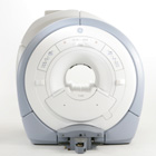
From GE Healthcare;
The GE Signa HDx MRI system is a whole body magnetic resonance scanner designed to support high resolution, high signal to noise ratio, and short scan times.
The 1.5T Signa HDx MR Systems is a modification of the currently marketed GE 1.5T machines, with the main difference being the change to the receive chain architecture that includes a thirty two independent receive channels, and allows for future expansion in 16 channel increments. The overall system has been improved with a simplified user interface
and a single 23" liquid crystal display, improved multi channel surface coil connectivity, and an improved image reconstruction architecture known as the Volume Recon Engine (VRE).
Device Information and Specification CLINICAL APPLICATION Whole body CONFIGURATION Compact short bore Standard: SE, IR, 2D/3D GRE and SPGR, Angiography: 2D/3D TOF, 2D/3D Phase Contrast; 2D/3D FSE, 2D/3D FGRE and FSPGR, SSFP, FLAIR, EPI, optional: 2D/3D Fiesta, FGRET, Spiral, Tensor, 2D 0.7 mm to 20 mm; 3D 0.1 mm to 5 mm 128x512 steps 32 phase encode POWER REQUIREMENTS 480 or 380/415 less than 0.03 L/hr liquid helium | | | | | |
| | | Searchterm 'SCan Time' was also found in the following services: | | | | |
| | |
| |
|

From GE Healthcare;
The Signa HDx MRI system is GE's leading edge whole body magnetic resonance scanner designed to support high resolution, high signal to noise ratio, and short scan times.
Signa HDx 3.0T offers new technologies like ultra-fast image reconstruction through the new XVRE recon engine, advancements in parallel imaging algorithms and the broadest range of premium applications. The HD applications, PROPELLER (high-quality brain imaging extremely resistant to motion artifacts), TRICKS (contrast-enhanced angiographic vascular lower leg imaging), VIBRANT (for breast MRI), LAVA (high resolution liver imaging with shorter breath holds and better organ coverage) and MR Echo (high-definition cardiac images in real time) offer unique capabilities.
Device Information and Specification CLINICAL APPLICATION Whole body
CONFIGURATION Compact short bore SE, IR, 2D/3D GRE, RF-spoiled GRE, 2DFGRE, 2DFSPGR, 3DFGRE, 3DFSPGR, 3DTOFGRE, 3DFSPGR, 2DFSE, 2DFSE-XL, 2DFSE-IR, T1-FLAIR, SSFSE, EPI, DW-EPI, BRAVO, Angiography: 2D/3D TOF, 2D/3D phase contrast vascular IMAGING MODES Single, multislice, volume study, fast scan, multi slab, cine, localizer H*W*D 240 x 2216,6 x 201,6 cm POWER REQUIREMENTS 480 or 380/415, 3 phase ||
COOLING SYSTEM TYPE Closed-loop water-cooled grad. | | | | | |
| | | | | |
| |
|
(SFP or SSFP) Steady state free precession is any field or gradient echo sequence in which a non-zero steady state develops for both components of magnetization (transverse and longitudinal) and also a condition where the TR is shorter than the T1 and T2 times of the tissue. If the RF pulses are close enough together, the MR signal will never completely decay, implying that the spins in the transverse plane never completely dephase.
The flip angle and the TR maintain the steady state. The flip angle should be 60-90° if the TR is 100 ms, if the TR is less than 100 ms, then the flip angle for steady state should be 45-60°.
Steady state free precession is also a method of MR excitation in which strings of RF pulses are applied rapidly and repeatedly with interpulse intervals short compared to both T1 and T2. Alternating the phases of the RF pulses by 180° can be useful. The signal reforms as an echo immediately before each RF pulse; immediately after the RF pulse there is additional signal from the FID produced by the pulse.
The strength of the FID will depend on the time between pulses (TR), the tissue and the flip angle of the pulse; the strength of the echo will additionally depend on the T2 of the tissue.
With the use of appropriate dephasing gradients, the signal can be observed as a frequency-encoded gradient echo either shortly before the RF pulse or after it; the signal immediately before the RF pulse will be more highly T2 weighted.
The signal immediately after the RF pulse (in a rapid series of RF pulses) will depend on T2 as well as T1, unless measures are taken to destroy signal refocusing and prevent the development of steady state free precession.
To avoid setting up a state of SSFP when using rapidly repeated excitation RF pulses, it may be necessary to spoil the phase coherence between excitations, e.g. with varying phase shifts or timing of the exciting RF pulses or varying spoiler gradient pulses between the excitations.
Steady state free precession imaging methods are quite sensitive to the
resonant frequency of the material.
Fluctuating equilibrium MR (see also FIESTA and DRIVE)and linear combination SSFP actually use this sensitivity for fat suppression. Fat saturated SSFP (FS-SSFP) use a more complex fat suppression scheme than FEMR or LCSSFP, but has a 40% lower scan time.
A new family of steady state free precession sequences use a balanced gradient, a gradient waveform, which will act on any stationary spin on resonance between 2 consecutive RF pulses and return it to the same phase it had before the gradients were applied.
This sequences include, e.g. Balanced Fast Field Echo - bFFE, Balanced Turbo Field Echo - bTFE, Fast Imaging with Steady Precession - TrueFISP and Balanced SARGE - BASG. See also FIESTA. | | | |
• View the DATABASE results for 'Steady State Free Precession' (20).
| | | | | | Further Reading: | News & More:
|
|
| |
| | | | |
| | |
| | | |
|
| |
 | Look
Ups |
| |


